Impact of Different Modeling Approaches in Field Focusing Predictions of Hyperthermia Cancer Therapy
The hyperthermia therapy (HT), i.e. an increase of the body tissues temperature to 39-43 °C caused by the exposure to non-ionizing electromagnetic radiation (like radiofrequency or microwaves), has been demonstrated to sensitize tumours to radiotherapy and chemotherapy (i.e. cancer therapies are more effective at the same dose), without adding toxicity. The fundamental aim of an optimized hyperthermia treatment consists in heating the tumour region to a temperature of about 43 °C, while keeping the heat in the surrounding healthy tissues in a tolerable range to avoid the presence of hot spots, especially in thermosensitive tissues (like cerebrum, cerebellum, spinal cord, etc.). With the aid of the COMSOL Multiphysics® simulation software, we reproduced a simplified version of a hyperthermia array applicator [1], which is basically an array of patch antennas specifically designed to treat deep-seated tumours in the head and neck (H&N) region, and we compared the SAR (Specific Absorption Rate) maps obtained focusing the electromagnetic heating on the tumour mass for three different models of the neck region.
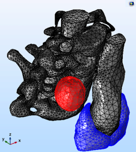
The first model consists in a very simple representation of the human neck, where the vertebras, the spinal cord, the trachea as well as the neck shape are modeled by means of cylinders, while the tumour is represented by a sphere. Then, we implemented a more realistic neck phantom importing in COMSOL Multiphysics® the 3D mesh models of the vertebras (from C3 to C7), the spinal cord and the laryngotracheal canal provided by the Visible Human Project (VHP-Female Version 2.2 [2]). In this second case, the tumour is modeled as a sphere of irregular shape and the thyroid gland is also reproduced. Finally, we used the “Interpolated Material Data” method [COMSOL Application ID: 59131] to import in COMSOL Multiphysics® the neck section extrapolated from the Zubal phantom voxel data [3]. For all the considered neck models, the electrical and thermal parameters have been properly assigned to the different tissues; then, by means of the COMSOL Multiphysics® RF and LiveLink™ for MATLAB® modules, an optimization of the array feedings was performed at a frequency of 434 MHz, to maximize the SAR on the tumour target volume, reducing the risk of hot-spots in the surrounding healthy regions. As a result, the impact of the different neck simulation approaches on the optimized SAR maps has been analyzed.